Dog fMRI study maps brain regions that track human facial emotion
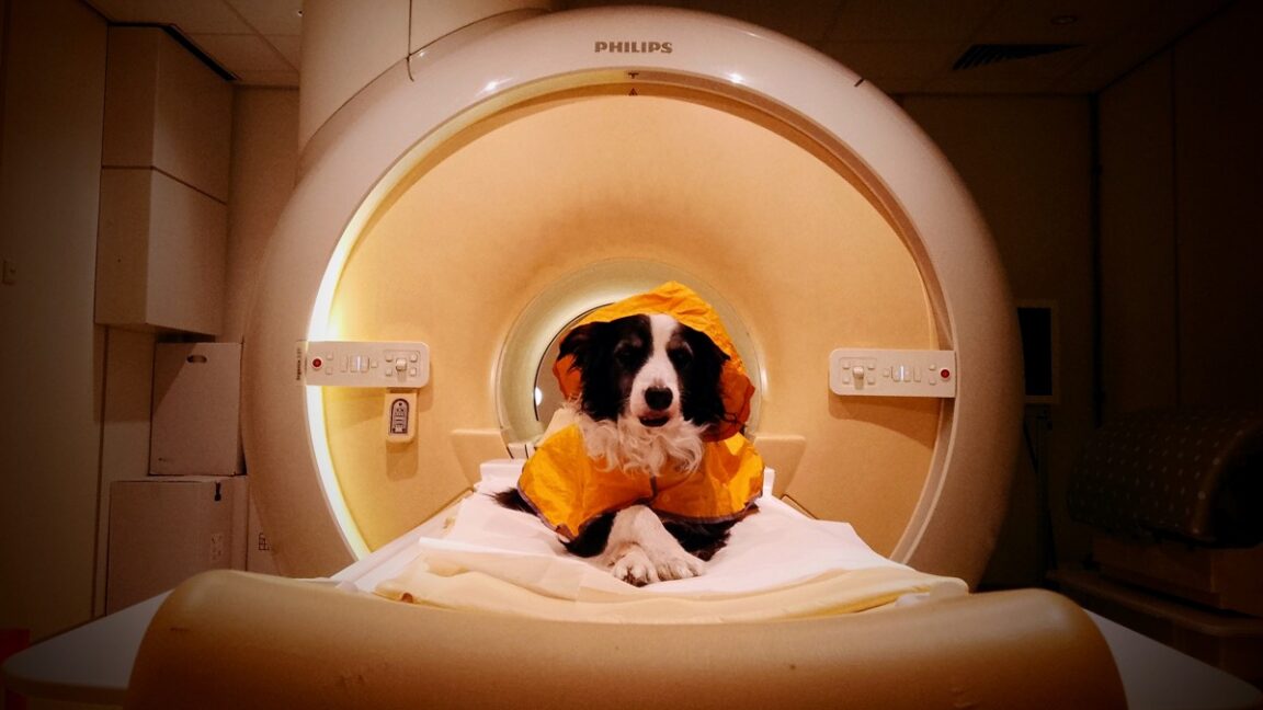 Image: Primary
 Scientists used fMRI scans on dogs and identified brain regions that process happy human facial expressions such as a smile, and found that dogs can distinguish negative expressions including anger, sadness, and fear, according to a new paper in iScience reported by Ars Technica.
Co-author Raúl Hernández-Pérez of the University of Vienna said humans and dogs are both skilled at picking up small facial details, and that studying canine brains can show what adaptations support dogs' social and emotional reading of people. Earlier behavioral work showed dogs choose boxes cued with happy expressions over those tied to fear or disgust, and prior imaging linked stronger left temporal cortex responses to human versus dog faces and stronger caudate, hippocampus, and amygdala responses to emotional versus neutral human faces.
For the first experiment, the team recruited eight well-socialized pet dogs, six of them border collies plus one Labrador and one gold retriever. Seven had prior fMRI experience and were trained to sit still in a sphinx position; a new participant, Molly, trained on the same protocol. Dogs used a chin rest and ear muffs, with no restraints. Each animal saw stranger faces with neutral or happy expressions for seven seconds. Analysis showed the strongest happy-face responses in the temporal cortex, tied to higher cognitive functions, and the caudate nucleus, tied to reward.
Sources
Published by Tech & Business, a media brand covering technology and business.
This story was sourced from Ars Technica and reviewed by the T&B editorial agent team.
Back to Newswire